HomeProductsRaman Spectroscopy SystemSEER Model Microscopic Imaging Raman Spectrometer
  • White-Light Microscopic Imaging Raman Spectroscopy System
  • White-Light Microscopic Imaging Raman Spectroscopy System

    SEER Model Microscopic Imaging Raman Spectrometer

    As a rapid, non-destructive analytical technique with unique "fingerprint" specificity, Raman spectroscopy directly reveals information about material chemical structure, phase and morphology, and molecular interactions, and is now widely used in physics and chemistry research at universities and scientific research institutions.

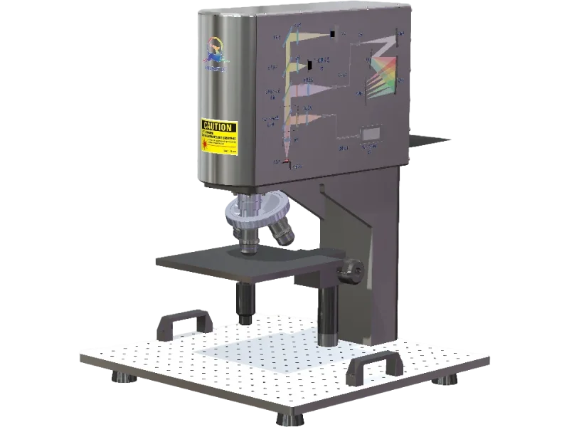
    The microscopic Raman instrument serves spectroscopy education with its semi-open structural design that breaks away from the conventional black box design of traditional Raman systems, making optical principles accessible and creating a new teaching platform for educators and students. This instrument provides high spectral resolution, detection sensitivity, and signal-to-noise ratio, along with multiple teaching functions including white light microscopic imaging and Raman spectroscopy detection.

    Product Features
    ・High SNR
    ・High Spatial Resolution
    ・Cost-Effective
    ・High Stability
    ・Built-in Raman Calibration
    ・Integrated Optical Path Diagram
    ・Lesson Plans & Training Videos
    Best For
    ・Mineral Analysis
    ・Pharmaceutical Testing
    ・Jadeite Authentication
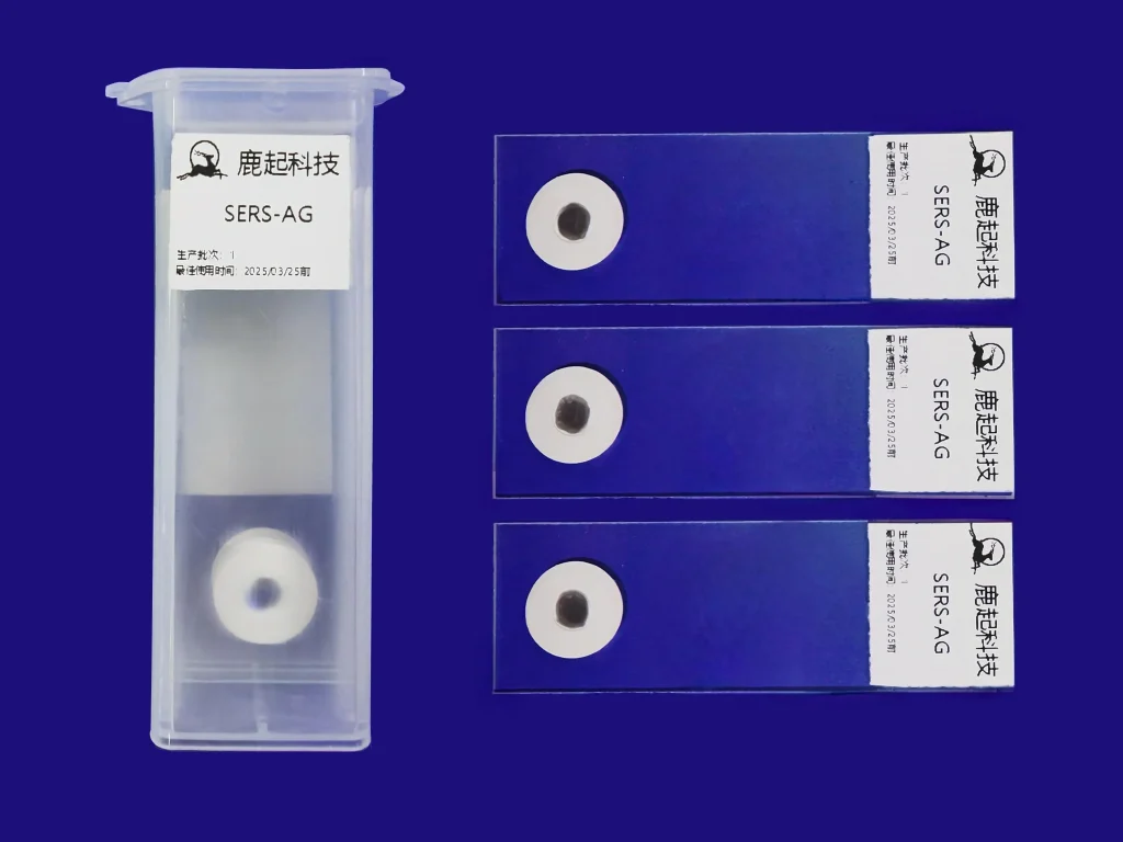
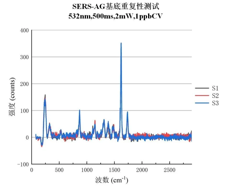
    ・SERS Detection
    ・Particle Characterization
    ・Research Applications
    ・Illicit Substance Detection
    ・Perovskite Solar Cell Inspection

HOW CAN WE HELP YOU

You can contact us any way that is convenientfor you. We are available 24/7 via email ortelephone.
+44 7496521078 sale@rudeeroptics.com
  • Product Details
  • Download
Specifictions
Model:  SEER Series
Spectrometer: Raman Spectral Range SEER-532nm:150cm^−1-3600cm^−1
SEER-785nm:200cm^−1-2500cm^−1
Spectral Resolution SEER-532: 7 cm⁻¹ (25 μm slit, measured)
SEER-785: 6 cm⁻¹ (25 μm slit, measured)
Wavenumber Accuracy ±3cm^−1
Detector: Integration Time 9.5ms-20s
SNR 600:1
Dynamic Range 79000:1
Laser: Excitation Wavelength 532 nm or 785 nm (optional)
Linewidth <0.1nm
Laser Power Multimode: 100 mW / 500 mW
Single-mode: 100 mW / 500 mW
Microscopic Imaging Spot Size 3 μm @100x (532 nm, measured)
Light Source LED white light illumination
Tube Lens Focal Length 180mm
Laser Spot Observation Simultaneous laser spot and microscopic image observation
Objective Lens 10x/20x/50x/100x (optional)
Software & Interface OS:32/64bit Windows 7/10/11
Interface:USB2.0
Software:RudeerTek RamanSoft

Typical Spectrum

SERS-AG
RudeerTek’s SERS substrate provides a trace molecular analysis solution based on Surface-Enhanced Raman Spectroscopy (SERS) technology, specifically designed for detection scenarios requiring ultra-high sensitivity.

WhatsApp

Leave a message!

Leave a message!

Please enable JavaScript in your browser to complete this form.